. 2020 Sep 24;9:e62514. doi: 10.7554/eLife.62514

Figure 4. Major ABD conformational rearrangements upon actin binding.

(A) Cartoons and crystal structures of pre-bound (meta)vinculin ABD (PDB 1st6, dark orange, top) and α-catenin ABD (PDB 4igg chain B, dark pink, bottom). Regions that undergo major rearrangements are highlighted in brown (metavinculin) or magenta (α-catenin); regions not resolved in the actin-bound cryo-EM structures are transparent. (B) Cryo-EM structures of metavinculin ABD (orange, top) and α-catenin ABD (pink, bottom). Flexible regions colored as in (A); actin, shades of blue. (C) Superimposed pre-bound and post-bound structures of metavinculin ABD (top) and α-catenin ABD (bottom). Green arrows indicate displacement of helices; rotation angles indicate repositioning of C-terminal extensions (CTEs).

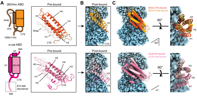
Figure 4.

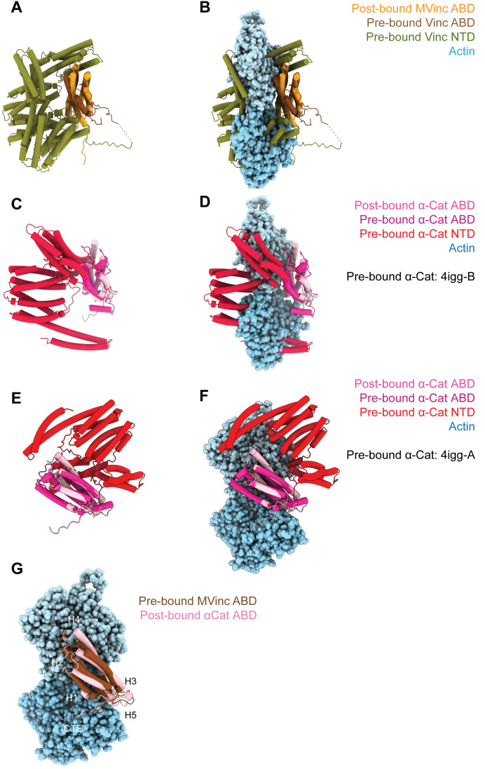
Figure 4—figure supplement 1. (Meta)vinculin and α-catenin activation mechanisms.

Figure 4—figure supplement 1.

(A and B) Superimposed pre-bound full-length vinculin crystal structure (PDB 1st6) and post-bound metavinculin ABD cryo-EM structure in the absence of actin (A), or in the presence of actin (B). (C, D, E, and F) Superimposed pre-bound full-length α-catenin crystal structures (PDB 4igg: C and D: Chain B; E and F: Chain A) and post-bound α-catenin ABD cryo-EM structure without (C and E) or with (D and F) actin. (G) Superimposed pre-bound full-length vinculin crystal structure (PDB 1st6) and post-bound α-catenin ABD cryo-EM structure in the presence of actin highlights the similar orientations of their CTEs.
Figure 4—video 1. Overall conformational changes in ABDs upon actin binding.
Download video file (246.7KB, mp4)
Linear interpolation morph of pre-bound to post-bound structures of the (meta)vinculin ABD (left) and α-catenin ABD (right). Colors, views, and superpositions are equivalent to Figure 4A and B.
Figure 4—video 2. Rearrangements in the helical bundle regions of ABDs upon actin binding.
Download video file (144.8KB, mp4)
Linear interpolation morph of pre-bound to post-bound structures of the (meta)vinculin ABD (left) and α-catenin ABD (right), focused on their similar twisting re-arrangements in helices H2–H5. Colors, views, and superpositions are equivalent to Figure 4C.