. 2020 Sep 24;9:e62514. doi: 10.7554/eLife.62514

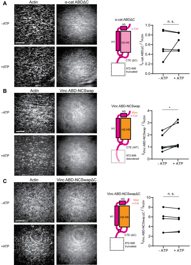
Figure 6. The distal tip of α-catenin’s C-terminus is a force detector.

(A, B, and C) TIRF force reconstitution assays. Left: Representative movie frames in the presence and absence of ATP. Scale bar, 30 μm. Right: Cartoon of ABD construct (left) and paired analysis of the overall intensity ratio change upon ATP addition (right). Wilcoxon signed-rank test: *p<0.05; n.s. (not significant), p>0.05. Constructs assayed were: α-cat ABDΔC (A) (αE-catenin664-871, N = 5, p=0.81); Vinc ABD-NCSwap (B) (αE-catenin664-708-vinculin916-1041-αE-catenin837-906, N = 6, p=0.031); Vinc ABD-NCSwapΔC (C) (αE-catenin664-708-vinculin916-1041-αE-catenin837-871, N = 5, p=0.63). Concentration of ABD constructs: 2 μM.

Figure 6.

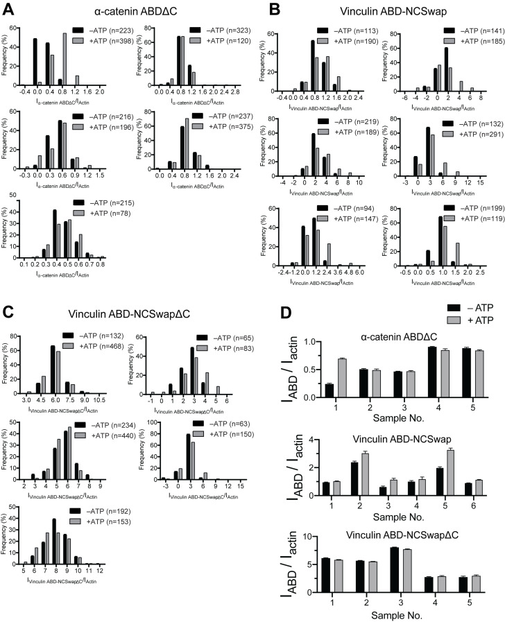
Figure 6—figure supplement 1. Intensity ratio distributions of single-filament regions for α-catenin and vinculin CTE mutants.

Figure 6—figure supplement 1.

(A, B, and C) Time-averaged IABD/Iactin intensity ratio distributions of single-filament regions before and after ATP addition for α-catenin ABDΔC (A); Vinc ABD-NCSwap (B); Vinc ABD-NCSwapΔC (C). These data were used for the analysis presented in Figure 6A–C. (D) Bar plots of mean ± SEM of high-force and low-force averages for all trials displayed in Figure 6.
Figure 6—video 1. TIRF force reconstitution assay of α-catenin ABDΔC truncation mutant.
Download video file (1MB, mp4)
First 100 s of representative dual-color TIRF-assay movies of actin (left) and α-catenin ABDΔC (right) in the absence (top) and presence (bottom) of ATP, as shown in Figure 6A. Scale bar, 20 µm. This construct loses force-activated binding activity, binding actin equivalently in both conditions.
Figure 6—video 2. TIRF force reconstitution assay of Vinc ABD-NCSwap chimera.
Download video file (796.7KB, mp4)
First 100 s of representative dual-color TIRF-assay movies of actin (left) and Vinc ABD-NCSwap (right) in the absence (top) and presence (bottom) of ATP, as shown in Figure 6B. Scale bar, 20 µm. This construct gains force-activated binding activity, displaying impaired binding in the absence of force.
Figure 6—video 3. TIRF force reconstitution assay of Vinc ABD-NCSwapΔC chimera truncation mutant.
Download video file (600.7KB, mp4)
First 100 s of representative dual-color TIRF-assay movies of actin (left) and Vinc ABD-NCSwapΔC (right) in the absence (top) and presence (bottom) of ATP, as shown in Figure 6C. Scale bar, 20 µm. This construct loses force-activated binding activity versus the Vinc ABD-NCSwap chimera, binding actin equivalently in both conditions.