
Fig. 3.
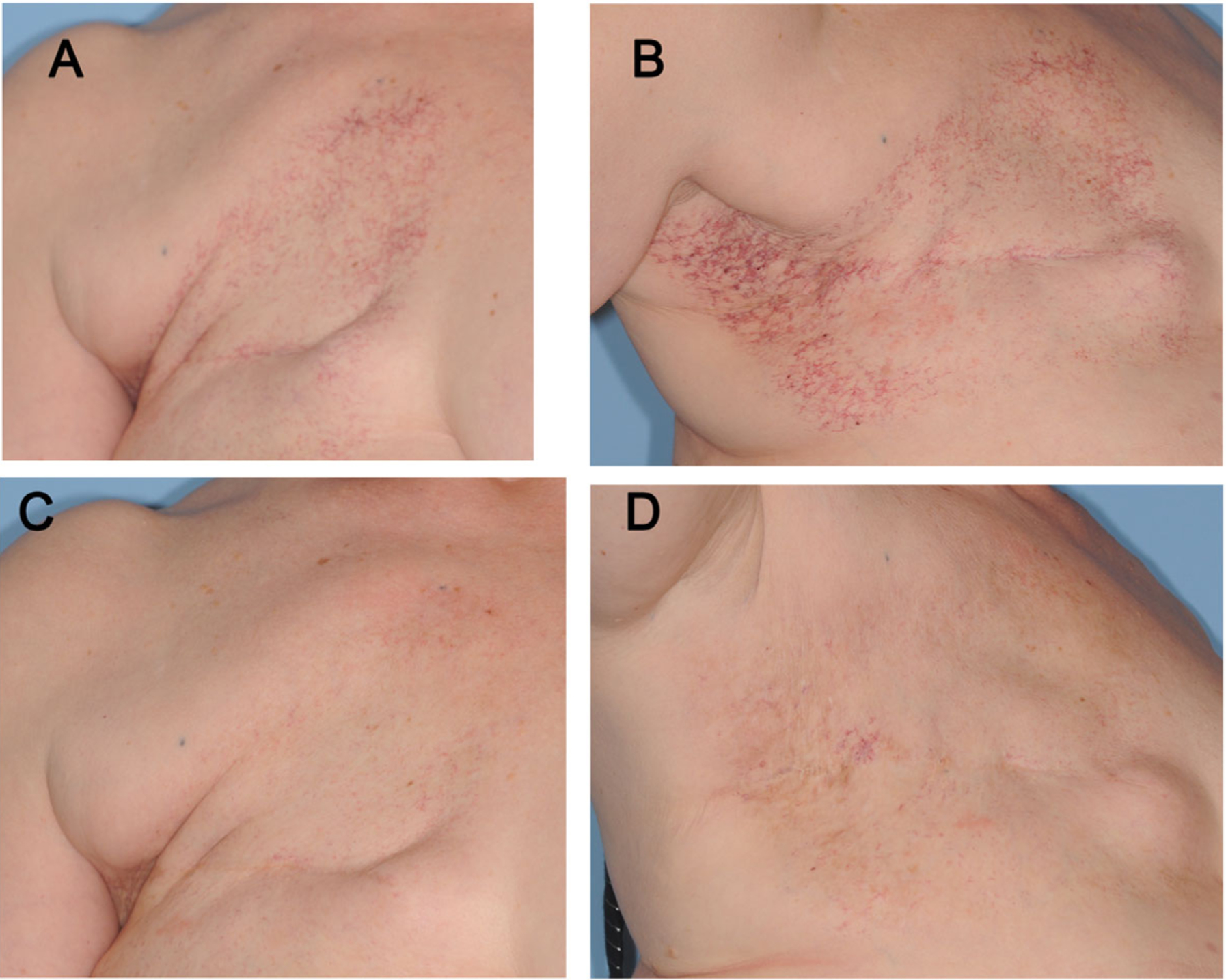
Clinical improvement in RIBT following treatment with the 595 nm PDL. (A) Pre-treatment RIBT across the chest wall and décolletage. (B) This patient’s RIBT also extended up into her axilla. (C) Frontal and (D) axillary views 4 weeks after treatment number two, with 75–100% clearance (10 mm spot size, average fluence 7 J/cm2, 3 milliseconds pulse duration). RIBT, radiation-induced breast telangiectasias; PDL, pulsed dye laser.