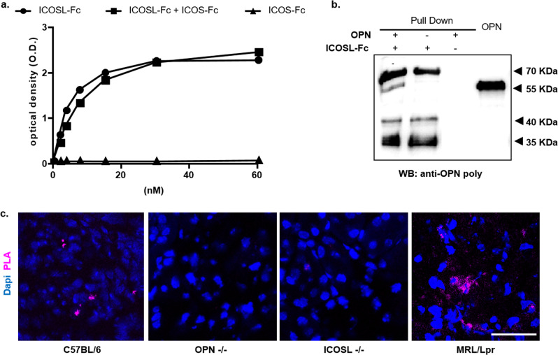
Fig. 2. OPN binds the extracellular portion of ICOSL.
a ELISA-based interaction assay. Recombinant OPN was used as capture protein on 96 well plates and the binding of titrated amounts of soluble recombinant ICOSL-Fc was evaluated alone (black circle) or mixed 1:1; with ICOS-Fc (black square) or of ICOS-Fc alone (black triangle). Data are expressed as means ± standard error (n = 3 technical replicate). b Pull-down assay. Schematic presentation of the pull-down assay used to detect ICOSL/OPN binding, in which ICOSL-Fc was used as a Sepharose-bound bait protein incubated (first lane) with OPN. The second (ICOSL-Fc) and third lanes (OPN) represent the negative controls in which only one of the two interactors were present. The membrane was blotted with an anti-OPN polyclonal antibody. A representative immunoblot of two independent experiment is shown (uncropped blot is shown in Supplementary Fig. 6). c Proximity Ligation Assay (PLA) in mice kidneys. PLA signal (violet) shows protein-protein interactions between ICOSL and OPN; C57BL/6J and MRL/Lpr chosen as positive controls, while OPN-/- and ICOSL-/- as negative ones. Four images at a magnification of ×40 were analyzed for each sample, considering 3 mice per group (n = 3 biological replicate).

