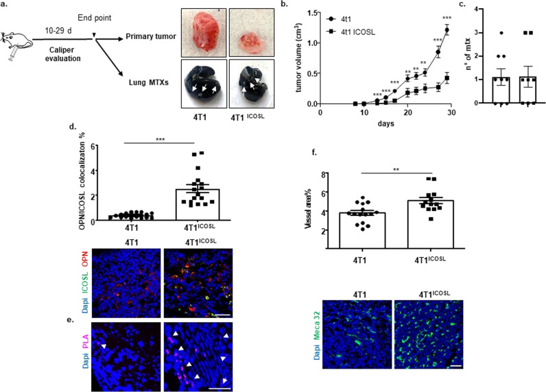
Fig. 8. OPN/ICOSL binding modulates tumor growth, angiogenesis, and metastatization in vivo.
a Schematic illustration of experimental in vivo approach and images of excised 4T1and 4TICOSL tumors, where white arrows indicate metastases. b Primary tumor growth of 4T1 cells in comparison with 4TICOSL cells. 0.1 × 10⁶ 4T1 and 4T1ICOSL cells were subcutaneously injected into the mammary fat pad of Balb/c mice (n = 8–9 biological replicate per group). Tumor growth was monitored using a caliper by measuring tumor size in 3 cross-directions. The Mann–Whitney test was used to compare differences in tumor growth between the two groups per day, **P < 0.01, ***P < 0.001. c Numbers of 4T1 and 4T1ICOSL tumor lung metastases at the end point (day 29). d OPN and ICOSL protein level and localization were evaluated by confocal analysis. Images were quantified using ImageJ software which permits to calculate the ratio between red (OPN) and green (ICOSL) channels; values are expressed as percentage of red-green co-staining. Dot plot shows OPN and ICOSL fluorescence intensity colocalization (3–4 biological replicate) and the Mann–Whitney test was used. e PLA on primary tumor specimens showing OPN–ICOSL interaction. f As assessed by confocal analysis of Meca32 immunostaining, vessel density in 4T1ICOSL tumors was significantly increased (by 26%) compared with wild-type 4T1. The percentage of surface area occupied by vessels was quantified as Meca32 positive staining. Results have been analyzed using unpaired Mann–Whitney U test. Scale bars: 50 μm (n = 3–4 biological replicate). Four images at a magnification of ×40 were analyzed for each sample, considering three mice per treatment group. All data are expressed as means ± standard error.

