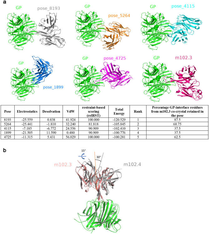
Figure 2.
Computational docking and mode of m102.4-GP binding. (a) The top five docked models identified by pyDockWeb along with co-crystal of m102.3 (PDB: 6CMG) are shown. The orientation of GP (green) is intact in all six panels. The table underneath shows the energetic terms along with percentage of GP-interface residues from m102.3 co-crystal that are retained in each of the five poses. (b) Structural superposition of m102.3 co-crystal (PDB: 6CMG) and pose_8193 using GP (green) as the reference. The angles that capture the differences in binding orientation are highlighted to show the differences. All figures were generated using PyMOL (https://www.pymol.org).

