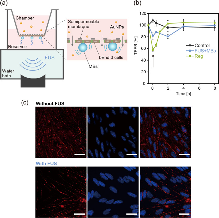
Figure 2.
In vitro investigation of BBB opening via the integration of a semipermeable culture insert and FUS exposure setup. (a) A schematic diagram of the apparatus constructed for evaluating the FUS-induced enhancement of BBB permeability to nanoparticles. (b) Time change in TEER induced by FUS exposure in the presence of MBs. The effect of treatment with 10 μM regadenoson (Reg) was also evaluated for comparison. The arrow indicates the time when FUS exposure or Reg treatment was conducted (defined as 0 h). The control group was the BBB model that received no treatment. N = 4. (c) Immunohistochemical staining of the tight junction-associated protein ZO-1 (red) in bEnd.3 cells, with and without FUS exposure, observed using a confocal laser scanning microscope. Cell nuclei were also stained with DAPI (blue). The scale bar represents 10 μm.

