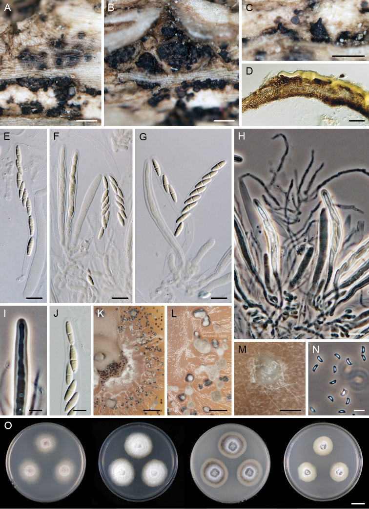
Figure 12.
Dendrophoma cytisporoides (CBS 144107). A–C ascomata D vertical section of the ascomal wall with remnants of the periderm E–G asci H paraphyses, asci and ascogenous hyphae I, J ascal apex K, L sterile primordia of sporodochia on MLA after 6 mo M sporodochium on OA after 10 wk N conidia O colonies on CMD, MLA, OA and PCA after 4 wk (from left to right). Scale bars: 300 μm (A–C, K); 20 μm (D); 10 μm (E–H); 5 μm (I, J, N); 100 μm (L, M); 1 cm (O).

