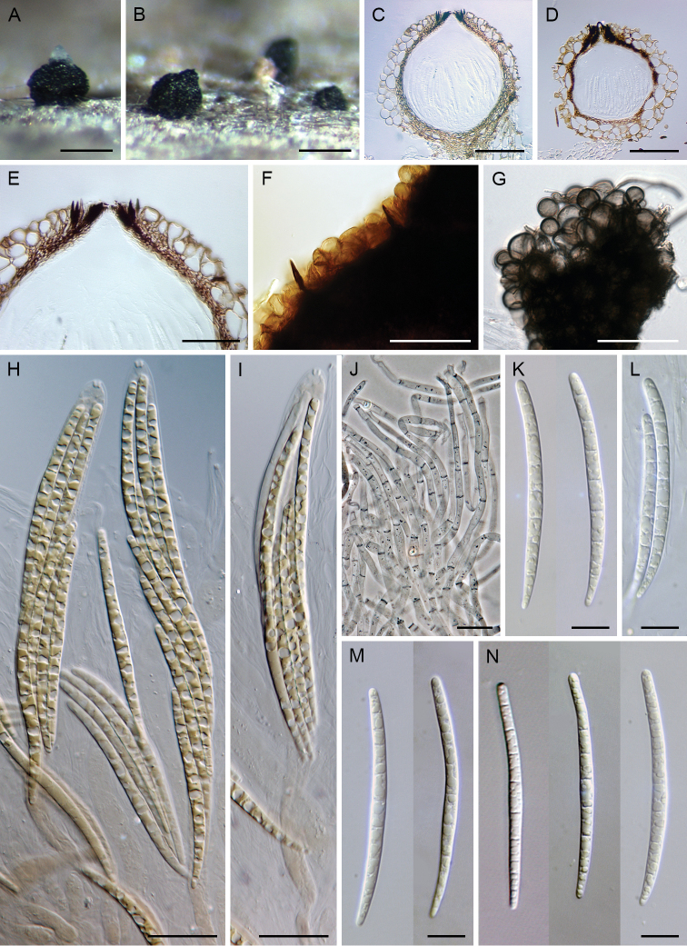
Figure 8.
Paragaeumannomyces longisporus. A, B ascomata C, D vertical section of ascomal wall E vertical section of ascomal wall and papilla with apical of setae F ascomal wall with setae G globose cells of the outer layer of the ascomal wall H, I asci J paraphyses K–N ascospores. Images: ILLS00121385 (A, B, G); S.M.H. 3860 (C, E, M); S.M.H. 2519 (D); ILLS00121386 (F, H–J, K); S.M.H. 2758 (L); S.M.H. 3809 (N). Scale bars: 250 μm (A–D); 50 μm (E–G); 20 μm (H–J); 10 μm (K–N).

