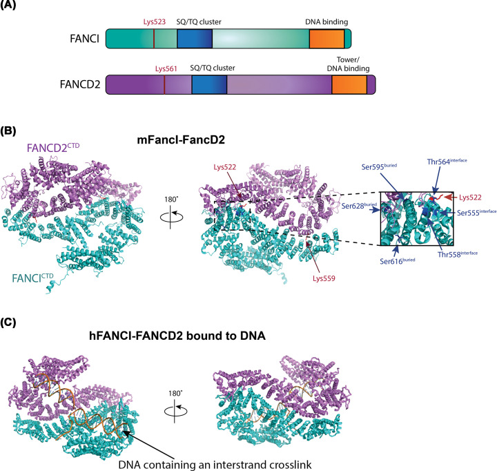
Figure 1. Overall structural comparisons of mFancI–FancD2 and hFANCI–FANCD2 bound to interstrand cross-linked DNA.
(A) The same domain architecture is shared by FANCI and FANCD2 and is shown schematically above. (B) Structure of mouse (m)FancI–FancD2 (PDB ID: 3S4W) where the ubiquitination sites, Lys559 residue of FANCD2 and Lys522 residue of FANCI are shown in red sticks; FANCI phosphorylation sites are shown in blue sticks. Close-up view of FANCI phosphorylation sites, Ser555, Thr558 and Thr564 exposed on the heterodimer interface, and Ser595, Ser616 and Ser628 residues buried in the complex. (C) Structure of human (h)FANCI–FANCD2 bound to interstrand cross-linked DNA (PDB ID: 6VAA). FANCD2 is colored in violet, FANCI in cyan and DNA in orange. Abbreviation: CTD, C-terminal domain.

