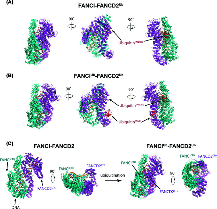
Figure 2. Overall structural comparison of FANCI–FANCD2, FANCI–FANCD2Ub and FANCIUb–FANCD2Ub.
(A) Structure of singly monoubiquitinated human FANCI–FANCD2Ub (PDB ID: 6VAF). (B) Structure of dually monoubiquitinated human FANCI Ub–FANCD2 Ub (PDB ID: 6VAE). (C) Comparison of FANCI–FANCD2 (PDB ID: 6VAA) and di-ubiquitinated FANCI–FANCD2 (PDB ID: 6VAE). FANCD2 is colored in violet, FANCI in cyan, ubiquitin in red and DNA in orange.

