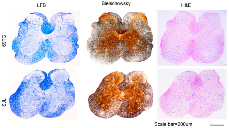
Figure 2.
Histology of the spinal cords from TCR-Tg and control mice infected with TMEV. Four different sections of the spinal cords of TCR-Tg and control SJL mice at 65 dpi were stained with Luxol Fast Blue (LFB), Bielschowsky silver staining or H&E. A representative sample is shown. Original magnification of the black scale bar = 200 μm.

