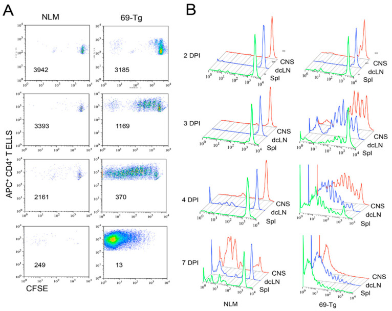
Figure 4.
Comparison of the initial proliferative responses of TCR-Tg and control CD4+ T cells in the CNS and periphery after TMEV infection. Negatively isolated CD4+ T cells from naive TCR-Tg and control SJL mice were labeled with CFSE and transferred intravenously into normal SJL mice. The recipient mice were infected intracerebrally with 1 × 10⁶ PFU TMEV. Cells from the recipients from the CNS, deep cervical lymph nodes (dcLN), and spleens were analyzed at 2, 3, 4, and 7 dpi for their cellular divisions using flow cytometry after staining with APC-labeled anti-CD4+ T cells in conjunction with CSFE intensity. (A) The division patterns of CFSE+CD4+ T cells in the spinal cords of the mice receiving either control or TCR-Tg CD4+ T cells. The number in each flow cytogram represents the mean CFSE intensity of the CD4+ T cells. (B) Comparison of the division patterns of the transferred CD4+ T cells in the CNS, dcLN, and spleen.

