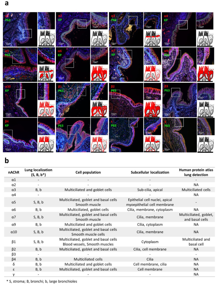
Figure 5.
nAChR localizations in human respiratory epithelia. (a) Representative micrographs showing the bronchial epithelia on formalin-fixed paraffin-embedded (FFPE) lung tissues stained for the nAChRs (all red), non-differentiated cells (p63 or pan-cytokeratin, green), and cell nuclei (DAPI, blue). Magnification corresponding to the selected area is shown. Drawings depict the localization of each nAChR subunit (in red). (b) Table summarizing nAChR subunit cellular and sub-cellular localization and the available microscopic data from the Human Protein Atlas (https://www.proteinatlas.org/). NA, not available; -, no detection.

