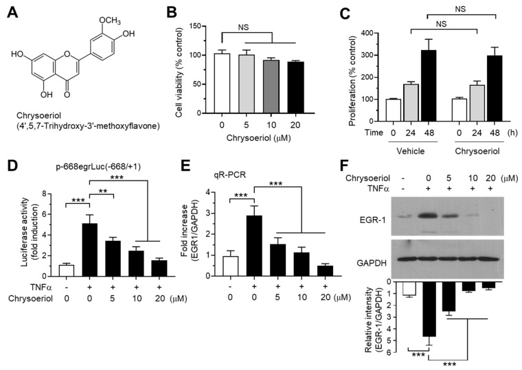
Figure 4.
Effect of chrysoeriol on EGR-1 expression. (A) Chemical structure of chrysoeriol. (B) Cell viability assay. MCF-7 cells were treated with vehicle (DMSO) or increasing concentrations of chrysoeriol (0–20 μM) for 24 h, and cell viability was determined using the Cell Counting Kit-8 (CCK-8). Graph bars represent the mean ± SD (n = 3). NS, not significant by Sidak’s multiple comparisons test. (C) MCF-7 cells were treated with vehicle (DMSO) or 20 μM chrysoeriol for different periods (0, 24, and 48 h), and cell proliferation was measured using an ELISA Colorimetric kit for the detection of 5-bromo-2′-deoxyuridine (BrdU) incorporation. Graph bars represent the mean ± SD (n = 3). NS, not significant by Sidak’s multiple comparisons test. (D) MCF-7 cells were transfected with 0.1 µg of EGR1 promoter–reporter p-668egrLuc(–668/ + 1). After 48 h, the cells were treated with 10 ng/mL TNFα in the presence or absence of chrysoeriol (0, 5, 10, 20 μM) for an additional 8 h, and the luciferase reporter activities were measured. (E,F) MCF-7 cells were pretreated with chrysoeriol (0, 5, 10, and 20 μM) for 30 min, followed by treatment with 10 ng/mL TNFα. After 1 h, cells were harvested and the EGR1 mRNA levels or protein levels were measured by qR-PCR (E) or immunoblot analysis (F), respectively. Band intensities were measured using the ImageJ software. Bars represent the mean ± SD (n = 3). ** p < 0.01, *** p < 0.001 by Dunnett’s multiple comparisons test. qR-PCR, quantitative real-time PCR.

