Figure 5.
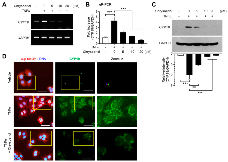
Effect of chrysoeriol on CYP19 expression. (A–C) MCF-7 cells were pretreated with chrysoeriol (0–20 μM) for 30 min, followed by treatment with 10 ng/mL TNFα. After 24 h, CYP19 expression was determined by RT-PCR (A), qR-PCR (B), and immunoblot analysis (C). GAPDH was used as an internal control. Band intensities were measured using the ImageJ software. Bars represent the mean ± SD (n = 3). ** p < 0.01, *** p < 0.001 by Dunnett’s multiple comparisons test. qR-PCR, quantitative real-time PCR. (D) MCF-7 cells cultured on coverslips were treated with 10 ng/mL TNFα in the absence or presence of 10 μM chrysoeriol, followed by fixation, permeabilization, and incubation with primary antibodies specific to α/β-tubulin and CYP19 aromatase for 2 h. After washing, Alexa Fluor 555 (for α/β-tubulin; red signal) and Alexa Fluor 488 (for CYP19; green signal) secondary antibodies were used to probe the samples for 30 min. Nuclear DNAs were stained with Hoechst 33258 for 10 min (blue signal). Fluorescently labeled cells were viewed under an EVOS FL fluorescence microscope. Nuclear DNA and α/β-tubulin were overlaid (left panels). Yellow boxed regions are zoomed-in on the far right. Scale bars represent 20 μm.

