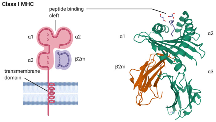
Figure 1.
A model depicting the major histocompatibility complex (MHC) class I HLA-B*27 heavy chain in complex with beta 2-macroglobulin (β2m) is shown on the left (created with BioRender.com). The alpha 1 (α1), alpha 2 (α2), and alpha 3 (α3) domains of the HLA-B*27 molecule and the peptide binding cleft composed of α1 and α2 domains of HLA-B*27 are indicated, as is the transmembrane domain inserted into the lipid bilayer of the plasma membrane. A ribbon model of the crystal structure of the human class I MHC molecule HLA-B*2705 heavy chain (green) bound to nona-peptide m9 (blue), and in complex with β2m (orange), is shown on the right. Orientation: cell surface at top of picture. The image was sourced from the Research Collaboratory for Structural Bioinformatics (RCSB; rcsb.org) Protein Data Bank (PDB) entry ID 1JGE (DOI:10.2210/pdb1JGE/pdb; [9]) generated by PISA [10].

