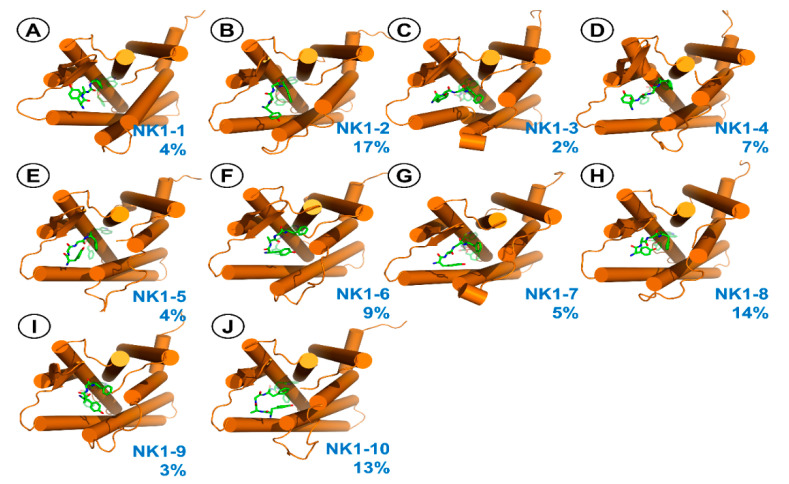
Figure 9.
Representative structures for the clusters from MD simulations of AA3266 in the NK1R binding site. Focus on the Tyr-d-Ala-Gly-Phe-fragment. (A) NK1-1 cluster, (B) NK1-2 cluster, (C) NK1-3 cluster, (D) NK1-4 cluster, (E) NK1-5 cluster, (F) NK1-6 cluster, (G) NK1-7 cluster, (H) NK1-8 cluster, (I) NK1-9 cluster, (J) NK1-10 cluster. Receptor is represented as orange cylinders (transmembrane helices, TM). The ligand is shown as green sticks. The blue number is the cluster population.

