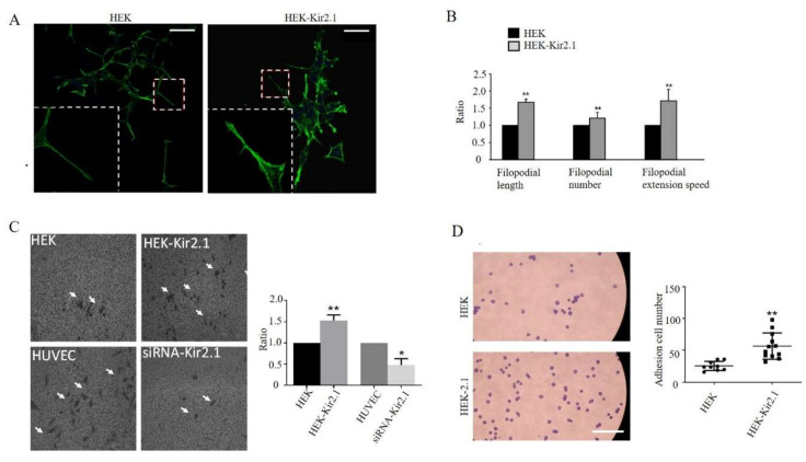
Figure 2.
Kir2.1 promotes filopodial extension, cell migration, and cell adhesion. (A) F-actin was visualized by FITC-conjugated phalloidin staining in HEK and HEK-Kir2.1 cell lines. The white dotted frames show the magnified images. Scale bar: 100 μm. (B) Quantification of filopodial length, filopodial number, and filopodial extension speed. ** p < 0.01. (C) Representative fields of the Transwell invasion assay under a phase-contrast microscope. Cells were grown, transfected, and then subjected to the Transwell assay. The white arrows point to the migrated cells stained by methylrosanilnium chloride solution. Quantitative results of the relative ratio of migrated cells is shown on the right; ** p < 0.01, * p < 0.05. (D) Representative fields of the adhesion assay. Cells were stained by methylrosanilnium chloride solution and measured by counting after adhesion for 45 min. scare bar: 500 µm. Quantification of total adhesion cell number per random field is shown on the right; ** p < 0.01.

