Figure 10.
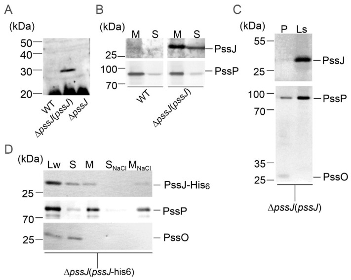
Localization of PssJ and PssJ-His6 proteins. (A–C) Western blotting with chicken anti-PssJ IgY affinity-purified antibodies to whole-cell lysates of the wild type strain, ΔpssJ(pssJ), and ΔpssJ (A); soluble (S) and membrane fractions (M) of the wild type strain and ΔpssJ(pssJ) (B); periplasmic proteins and lysate from the spheroplasts of ΔpssJ(pssJ) (C), Western blotting with mouse anti-His6 performed to whole cell lysate (Lw), soluble (S), membrane-containing (M), membrane-associated (SNaCl), and integral membrane (MNaCl) protein fractions of the ΔpssJ(pssJ-his6) strain (D). Secondary antibodies conjugated with horseradish peroxidase were used, which was followed by chemiluminescent detection (A). PssO is a soluble periplasmic protein and PssP is a typical transmembrane protein with two TMS. Both served as fraction purity markers. Loading was standardized to the volume of lysate, allowing visual assessment of the cellular distribution of PssJ.

