
Figure 3.
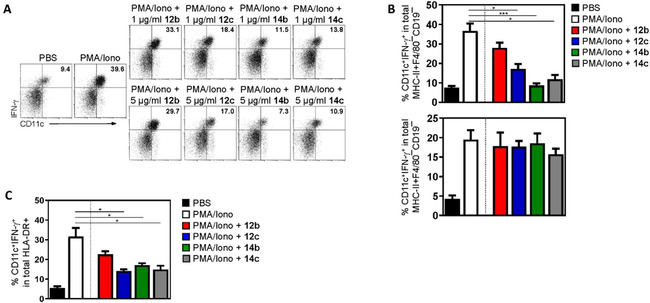
Compounds 12c, 14b and 14c prevented DC maturation and activation. A), B) Mouse DCs were generated from bone marrow precursors in the presence of GM‐CSF and IL‐4, activated with PMA and ionomycin (PMA/Iono) for 12 h, and stimulated with 12b, 12c, 14b or 14c in a concentration of 1 or 5 μg/mL for an additional 48 h. Control cells received an equal amount of PBS. Representative dot‐plots (A) and percentages of cells expressing the maturation marker CD11c as well as the pro‐inflammatory cytokine IFN‐γ after stimulation with 5 μg/mL of the KOR agonists (B) are shown. Data from n=4 wild‐type (top) or n=4 KOR‐deficient mice (bottom) are depicted. Cells are gated on MHC−II+F4/80−CD19− DCs, and IFN‐γ staining was performed after cell permeabilization. Data are presented as mean ±SD; * p<0.05 and *** p<0.01 vs. PMA/Iono treatment. C) Human DCs were purified from the peripheral blood of healthy donors and stimulated with PMA/Iono for 12 h. Subsequently, cells were incubated with compounds 12b, 12c, 14b, and 14c (5 μM/mL each) for an additional 48 h or received an equal amount of PBS. The percentages of cells expressing CD11c and IFN‐γ from n=4 healthy human donors are shown. Cells are gated on HLA‐DR, and IFN‐γ staining was performed after cell permeabilization. Data are presented as mean ±SD; * p<0.05 vs. PMA/Iono stimulation.