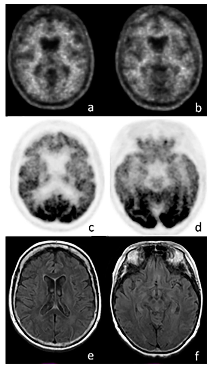
Figure 3.
Axial images of a PET scan performed with 18F-FBB (a,b), 18F-FDG (c,d), and MRI (e,f) in a patient with suspected frontotemporal dementia (FTD). The images show no amyloid burden in the cortex, while a significant reduction in cortical glucose consumption is visible in the frontal (c) and temporal lobes (d). MRI does not show any abnormal findings.

