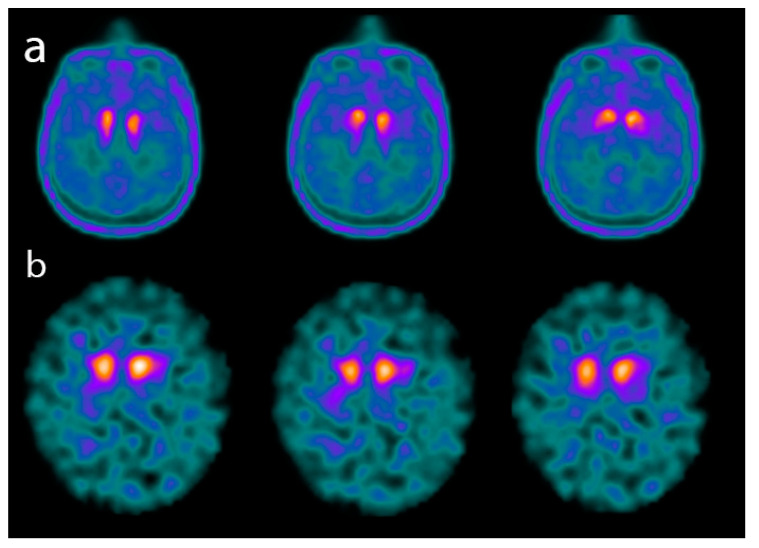
Figure 4.
Axial images of a PET scan performed with 18F-Fluoro-L-dopa (FDOPA) (a) and of a single-photon emission CT (SPECT) scan performed with 123I-FPCIT (DATSCAN) (b) in two patients affected by advanced dementia with Lewy bodies (DLB). The images show a significant reduction in both 18F-FDOPA and 123I-FPCIT uptake in the basal ganglia.

