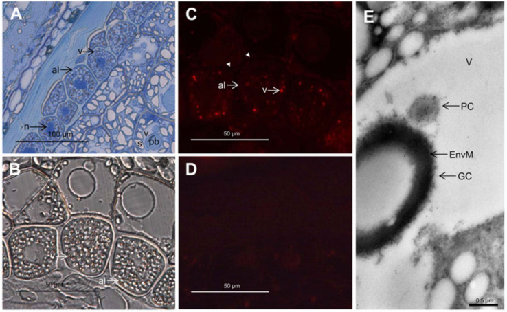
Figure 2.
Light (A–D) and immunoelectron (E) microscopy analysis of the localization of PAPhy in the developing wheat grain, approximately 18 days post anthesis. (A) Toluidine blue-stained semithin cross-section of endosperm, aleurone, and pericarp tissues. (B) Differential interference contrast microscopy with indications of the PSVs. (C) Immunofluorescence detection of PAPhy in 1 µm thick sections. The aleurone vacuoles are clearly labeled, while there is no fluorescence from any other compartment of the cell, the apoplast (arrowheads), or other cell types. (D) Immunofluorescence of a 1 µm thick section incubated with secondary antibody only. There is virtually no background from the secondary antibody. (E) Immunoelectron microscopy analysis showing an aleurone PSV with gold labeling of the protein crystalloid. al, Aleurone; EnvM, globoid enveloping membrane; GC, globoid crystal; n, nucleus; pb, protein body; PC, protein crystalloid; s, starch; v, vacuole. Reprinted in part from [25]. Copyright: American Society of Plant Biologists; http://www.plantphysiol.org.

