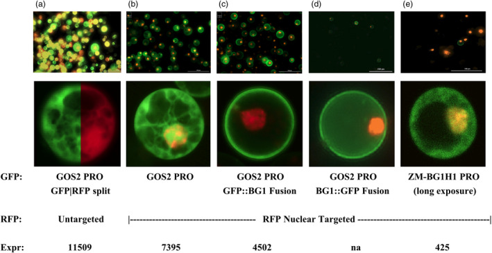
Figure 7.

Subcellular Localization of ZM‐BG1H1 Protein and Ectopic Expression Impact. a‐e. microscopic images of protoplast transfected with various Promoter::GFP reporter gene fusions as labelled at bottom of figure. Upper set of panels show lower magnification field with multiple protoplasts in view, and lower set of panels shows single close‐up view of a representative protoplast. Most protoplast ranged 20–30 μm diameter. Green colour emanates from the GFP reporter gene, and red colour from the RFP reporter gene. Panel A lower panel, subcellular localization control. Split image of a single protoplast, with GFP (left) and RFP (right) division, revealing broadly distributed GFP and RFP, respectively, in the cell. Panels b‐e, the RFP reddish‐orange colour restricted to the nucleus using nuclear localization signals. Panels a, b and e, the GFP is broadly distributed in the cell; panels c and d, the GFP is preferentially located at the protoplast plasma membrane.
