Figure 4.
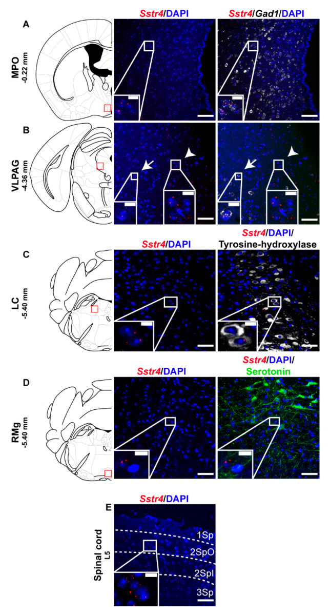
Representative Sstr4 expression in the descending anti-nociceptive system of WT mice. Sstr4 mRNA (red) counterstained with DAPI are shown in the medial preoptic area (MPO, Bregma −0.22 mm, (A)), in the ventrolateral periaqueductal gray matter (VLPAG, Bregma −4.36 mm, (B), arrow marks Sstr4 co-localization with Gad1, arrowhead points to Gad1-negative Sstr4 expressing neurons), in the locus coeruleus (LC, Bregma −5.40 mm, (C)) co-localized with tyrosine-hydroxylase (TH)-immunoreactive noradrenergic cells (white) and in the raphe magnus nucleus (RMg, Bregma −5.40 mm, (D)) where Sstr4 does not co-localize with serotoninergic cells (green). Panel (E) represents Sstr4 mRNA expressing cells in the superficial layers (I–III) of the spinal dorsal horn. (N = 2). Scale bar: 50 µm, inset scale bar: 10 µm.

