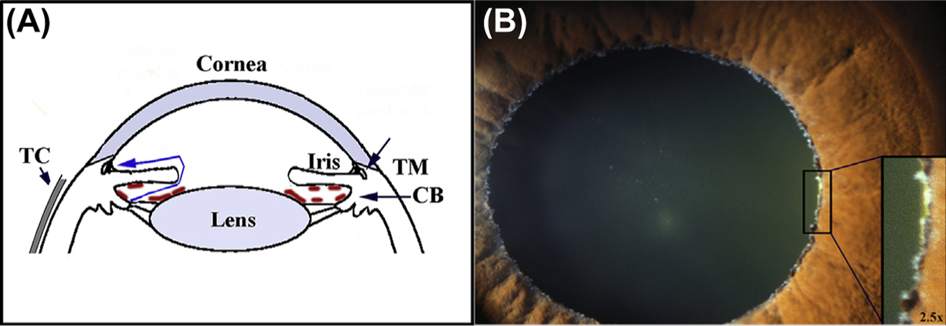
Fig. 1. (A) Schematic description of the cellular components of the anterior segments of the eye in relation to aqueous flow.

Fluid (blue line) is generated at the inner surface of the epithelium of the ciliary body CB) in the posterior aqueous chamber. From there it flows through the space between the iris and lens into the anterior aqueous chamber to exit the eye through the trabecular meshwork (TM) also referred to as the outflow facility. Increased, abnormal flow resistance of the facility causes elevation of the intraocular pressure (IOP). The increased pressure lead to the development of glaucoma, loss of retinal visual function due to retinal neural death. In individuals affected by exfoliation syndrome (XSF), proteinaceous material (shown tan marks in sketch) forms or deposits on the surface of the epithelial facing the posterior aqueous chamber, including the equator of the lens. The deposition is frequently associated with a type of glaucoma, exfoliation glaucoma, characterized by drug-unresponsive IOP increases and rapid progression of glaucoma. The Tenon’s capsule (TC) is a thin, elastic collagenous lining covering the sclera. (B) Flaky white deposits in the equator of the lens. The pupil has been dilated.
