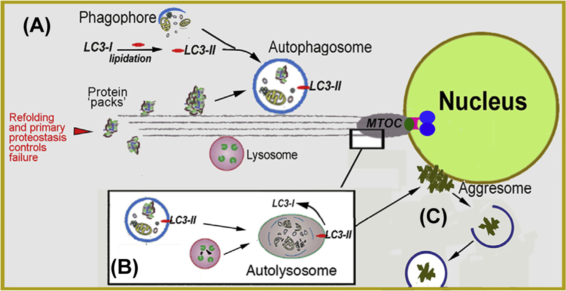
Fig. 5. Macroautophagy.

(A) Biogenesis of autophagosomes from precursor phagophores within the cytosol is orchestrated by the lipidated form of LC3 (LC3-II). Autophagosomes engulf ‘macroscopic’ cellular detritus, naturally decaying organelles, in particular, mitochondria (i.e., mitophagy) and misfolded or denatured proteins that have resisted the primary protein folding quality control mechanisms. (B) Autophagosomes fuse with lysosomes at the microtubule organizing center (MTOC). (C) Detritus that resist the combined proteostatic processes may accumulate in body inclusions, (e.g., Lewy body in senile dementia) categorized alternatively as aggresomes. If as demonstrated for tau protein propagation in Alzheimer’s and a-synuclein protein in Parkinson’s, then the aggregates may propagate to adjacent cells by particulate phagocytosis or endocytosis.
