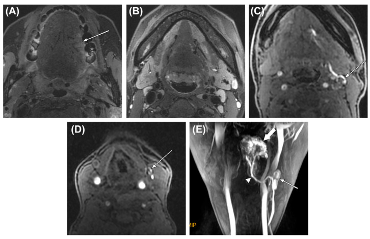
Figure 3.
A 38-year-old woman with oral tongue cancer and palpably negative neck. (A,B) Fat-saturated T2-weighted MRI scans show a shallow infiltrative tumor on the left lateral surface of oral tongue (arrow) and several small lymph nodes in the submandibular areas. (C,D) After peritumoral injection of contrast, MR lymphography revealed two first-enhanced lymph nodes in left level IB and IIA (arrows) on the first phase of the dynamic scan, respectively. (E) The maximum intensity projection reconstruction image of MR lymphography shows the contrast injection site in the tongue (thick arrow), the assumed sentinel lymph node (thin arrow), and the lymph vessel connecting them (arrowhead). After neck dissection, the assumed sentinel lymph nodes observed on MR lymphography revealed no metastasis on histologic examination [22]. Figure used with permission of John Wiley and Sons©, permission license number 4807630108259.

