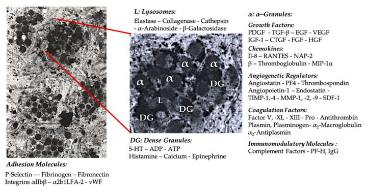
Figure 2.
Electron microscopic picture of a cluster of platelets from a PRP vial and a extrapolation of a single platelet (original magnification × 10,000) (from volunteer PE), representing the most familiar cellular constituents of α-granules (α), dense granules (DG), and lysosomes (L), including some platelet surface adhesion molecules. Adapted and modified from Everts et al. [61].

