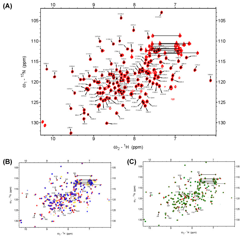
Figure 2.
The backbone assignment of DUSP22_WT and the superimposed 1H-,15N-HSQC spectra of the WT, S93N, N128A, and D57N. (A) The backbone amide resonance of residues in the WT is assigned on the 1H-,15N-HSQC spectrum. It exhibited 139 backbone amide resonances of 150 expected resonances (5 prolines removed) in DUSP22 and showed 93% completion. (B) The 1H-,15N-HSQC spectrum of the WT (red) was superimposed with the spectra of S93N (yellow) and N128A (blue). The spectra indicated that the mutants critically perturbed the solution structures and shifted several residues. (C) The 1H-,15N-HSQC spectrum of the WT (red) was superimposed with the spectra of D57N (green), which revealed the region perturbed by the DPN–triloop interaction. The labeled amino acids on the spectra were the residues in the D-loop, P-loop, and N-loop of the WT.

