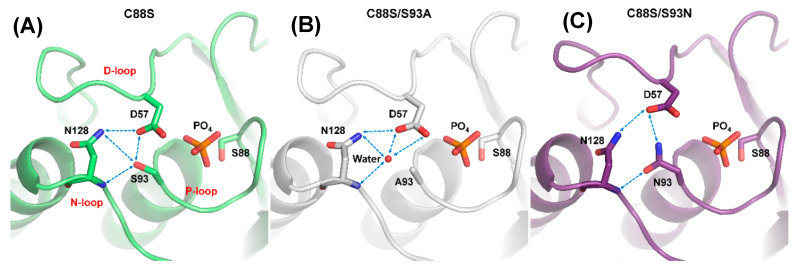
Figure 4.
Disruption of DPN–triloop interaction in the crystal structures of S93 mutants. (A–C) The crystal structures of C88S (green), C88S/S93A (white), and C88S/S93N (purple) indicated how the mutation affected the hydrogen bonding network. The hydrogen bonds are shown by blue dotted lines, and a water molecule is shown as a red ball. (A) The crystal structure of C88S was prepared to compare with the S93 mutants, and the DPN–triloop interaction was unchanged. (B) S93 was replaced with alanine, and the structure was similar to that of C88S. A water molecule (W61) participated in forming the hydrogen bonding with the side chain of D57, the side chain of N128, and the backbone amide of N128. (C) S93 was replaced with a somatic mutant, asparagine. N93 formed the hydrogen bonding with the side chain of D57 and the backbone amide of N128.

