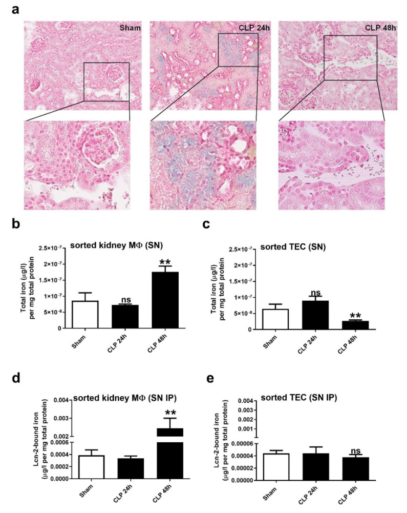
Figure 4.
Iron distribution in renal tissue during CLP-induced sepsis. (a) Iron accumulation in renal tissue restricted to the lumen of kidney tubules (blue colored areas) at 24 h after CLP detected by Perls’ staining. At the 48 h time-point, only brownish-stained hemosiderin deposits can be observed, which are mainly found in infiltrates. Pictures are representative for 5 animals in each group. (b,c) Measurements of total iron amounts in the supernatants of short-term cultivated and freshly isolated (b) renal MΦ and (c) TEC using AAS. (d,e) Lcn-2 was immunoprecipitated in the supernatants of short-term cultivated and freshly isolated (d) renal MΦ and (e) TEC, and Lcn-2-bound iron was quantified by AAS. For (b–e) n = 6 isolated primary peritoneal MΦ, TEC, and renal MΦ per group, ** p < 0.01; (b–d): one-way ANOVA followed by Tukey’s multiple comparison test. (e): Kruskal-Wallis test followed by Dunn’s post-hoc test.

