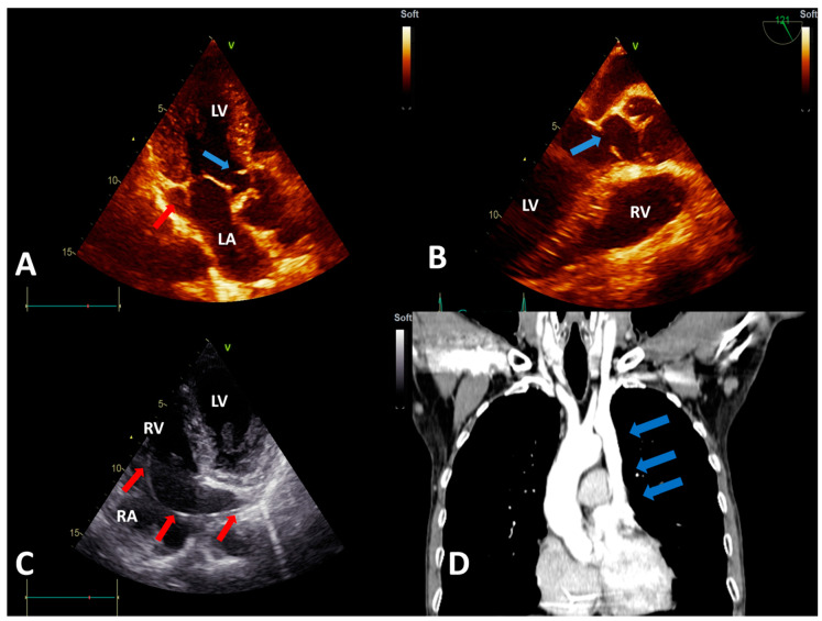
Figure 2.
(A) Transthoracic echocardiography (TTE), apical 3-chamber view showing the subaortic membrane (blue arrow) and the dilated coronary sinus (red arrow). (B) Transesophageal echocardiography view showing the subaortic membrane (blue arrow). (C) TTE, modified apical 4-chamber view showing the pacemaker lead (red arrows) entering the right atrium via the dilated coronary sinus (visualized in longitudinal section). (D) Computed tomography angiography showing the persistent left superior vena cava (blue arrows), with an absent right superior vena cava. Abbreviations: LA, left atrium; LV, left ventricle; RA, right atrium; and RV, right ventricle.

