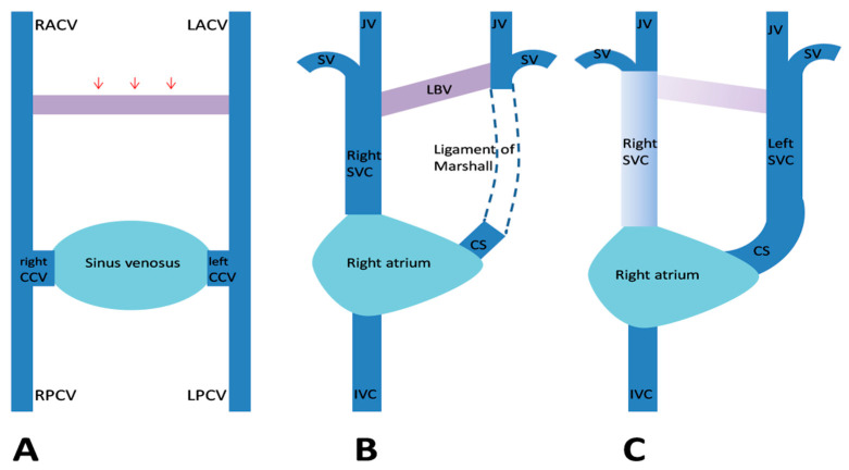
Figure 3.
Embryological development of a persistent left superior vena cava (SVC). (A) The venous drainage system of the embryo, with transverse anastomosis (red arrows) forming between the anterior cardinal veins. (B) Normal regression of the proximal segment of the left anterior cardinal vein (LACV) and the formation of the ligament of Marshall. (C) When this obliteration fails to occur, a left SVC develops. Abbreviations: CCV, common cardinal vein; CS, coronary sinus; IVC, inferior vena cava; JV, internal jugular vein; LBV, left brachiocephalic vein; LPCV, left posterior cardinal vein; RACV, right anterior cardinal vein; RPCV, right posterior cardinal vein; and SV, subclavian vein (Figure modified from Figure 5 in [17]).

