Figure 6.
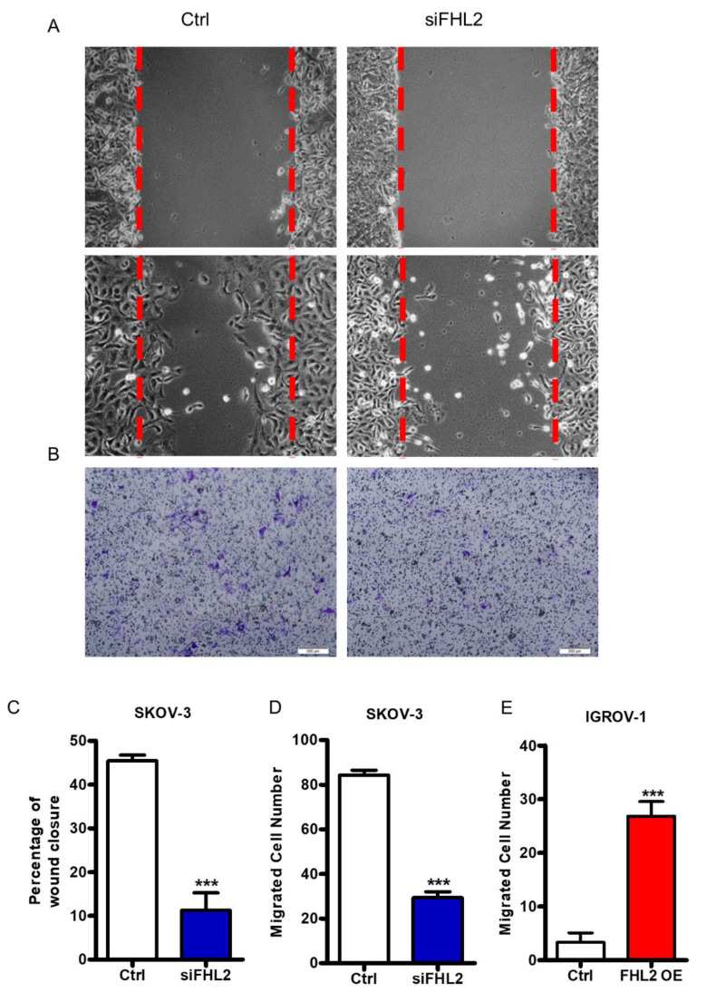
FHL2 regulates EOC cells migration and invasion. (A) Wound healing assay was performed to determine the effect of FHL2 on SKOV-3 cell migration. Representative images showed the migration of control cells (Ctrl, left panel) and FHL2 knockdown cells (siFHL2, right panel). Cells were allowed to migrate for 15 h before wound closure area measured. (B) Cell migration were analyzed by transwell migration assay. Representative images showed the migration of SKOV–3 control cells (left) and FHL2 knockdown cells (right). Scale bar: 200 μm. (C) The wound area was quantified with MicrosuitTM FIVE software. Each bar represents mean ± SEM. ***: p < 0.001 compared with control (Ctrl). (D) Migrated cells were counted manually under a microscope. Each bar represents mean ± SEM. ***: p < 0.001, compared with control. (E) Cell migration were evaluated by transwell assay in IGROV-1 control (Ctrl) and IGROV–1 FHL2 overexpression cells (FHL2 O/E). Each bar represents mean ± SEM. ***: p < 0.001.

