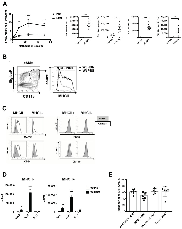
Figure 1.
Allergic asthmatic inflammatory conditions drive a heterogenous expression of MHCII in tissue resident alveolar macrophages. (A) Characterization of the allergic asthma phenotype of mice exposed to house dust mite extract (HDM) compared to non-asthmatic control (PBS). Upon HDM exposure, mice show increased airway hyperresponsiveness (AHR) upon methacoline exposure, as shown by increased airway resistance and strong inflammatory cell influx consisting mostly in eosinophils, T cells and to a lower extent neutrophils. Data show mean value ± SEM; n = 7–8 per group. Statistical significance in the AHR dose response curves was assessed using a two ways ANOVA. Statistical significance of the cellular recruitments in the airways was assessed using a t-test. ** p < 0.01; *** p < 0.001. (B) CD11c+SiglecF+ resident tissue-associated alveolar macrophages (tAMs) express different levels of MHCII complex upon HDM-driven allergic asthma inflammation. (C) Flow cytometric assessment of different markers of MHCII+ and MHCII− tAMs of HDM-treated mice, based on the gating provided in (B). Histograms show FMO control (grey) and MHCII signal (black) in the two subpopulations. The markers tested at the surface of CD11c+SiglecF+ tAMs were MerTK, CD64 and classical macrophages surface markers such as CD11b and F4/80. (D) RT-PCR analysis of MHCII+ and MCHII− tAMs for mRNA levels of Nos2, Arg1 and Ccr2 upon allergic inflammation. Data show mean value ± SEM of mRNA abundance reported to S14 mRNA in sorted MHCII+ and MCHII− tAMs; n = 4–7 per group. Statistical significance between PBS and HDM samples was assessed using a t-test; * p < 0.05; ** p < 0.01; *** p < 0.001. (E) Frequency of MHCII+ AMs in lungs of C57BL/6 WT and Ccr2−/− mice upon HDM-driven allergic asthma. Data show mean value ± SEM; n = 5–6 animals.

