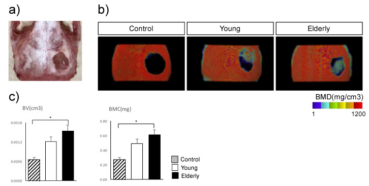
Figure 5.
Radiological findings of mouse calvarial defects 8 weeks after transplantation with cell sheets of TH-induced dental pulp stem cells (DPSCs) from young and elderly patients. (a) TH-induced DPSC sheets were cultured on temperature-responsive dishes for 14 days and then transplanted into mouse calvarial defects (3.5 mm in diameter). Eight weeks after transplantation, we dissected the calvaria (n = 4). (b) Micro-computed tomography images indicated the bone mineral density (BMD) values. The experiments were repeated three times, and one representative image is shown. (c) Quantification of bone volume (BV) and bone mineral content (BMC) of a 5 × 5 × 3 mm3 cuboid area in the center of a circular defect. Data are expressed as means ± standard deviation of five mice per group. * p < 0.05 by Student’s unpaired two-tailed t-test.

