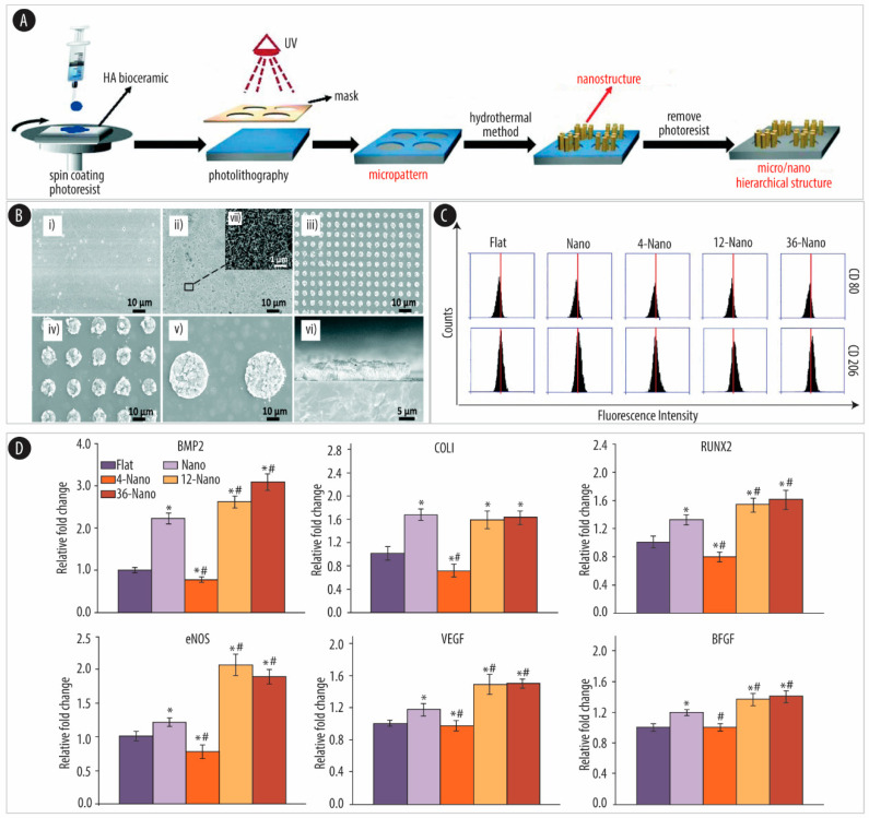
Figure 3.
Micro/nano hierarchical hydroxyapatite (HA) stimulation of osteogenetic and angiogenetic differentiation via macrophage immunomodulation. (A) Schematic of the preparation of HA bioceramics with micro/nano hierarchical surfaces using photolithography and hydrothermal techniques. (B) SEM images of HA bioceramics with different sized micro/nano hierarchical structures. (i) Control sample with a flat surface (Flat group). (ii) Only nanoneedle surface (Nano-group). (iii) 4 µm micro-dot/nanoneedle hierarchical surface (4-Nano group). (iv) 12 μm micro-dot/nanoneedle hierarchical surface (12-Nano group). (v) 36 µm micro-dot/nanoneedle hierarchical surface (36-Nano group). (vi) Cross-section of the 36-Nano group. (vii) The corresponding high magnification image of the 36-Nano-group. (scale bar: 10 µm (i, ii, iii, iv, v), 5 µm (vi), 1 µm (vii)). (C) Fluorescence intensity of cluster of differentiation (CD)80 (top) and CD206 (bottom) of RAW 264.7 (mouse mononuclear macrophages) cells on HA bioceramics with different topographical surfaces. (D) RT-qPCR results of the osteogenic gene expression (bone morphogenetic protein 2 (BMP-2), collagen type I (COLI), and runt-related transcription factor 2 (RUNX2)) of hBM-MSCs and angiogenic gene expression (endothelial nitric oxide synthase (eNOS), vascular endothelial growth factor (VEGF), and vascular endothelial growth factor (BFGF)) of human umbilical vein endothelial cells (HUVECs) cultured in RAW 264.7 cell-conditioned medium. * indicates significant difference (p < 0.05) compared to the Flat group. # indicates a significant difference (p < 0.05) compared to the Nano group. Adapted from [127] with permission from the Royal Society of Chemistry, 2019.

