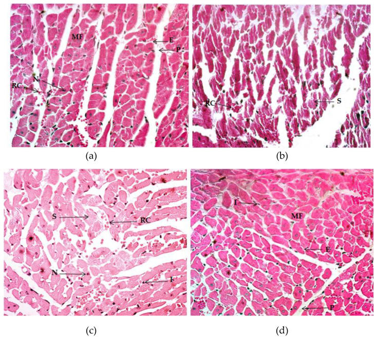
Figure 4.
Histology of the myocardium, Hematoxylin and Eosin (H&E) stain (40×). (a) Image of the control group. (b) Image of the exercise-training group without supplementation. (c) Image of the exercise-training groups plus vitamin C. (d) Image of the exercise-training group plus silymarin. MF: muscle fiber; E: endocardium; P: pericardium; RC: red cells; N: nuclei; S: splitting; I: inflammation.

