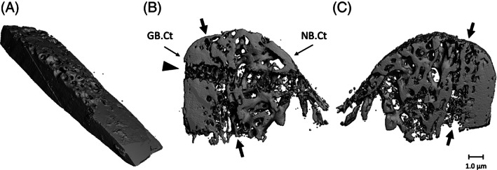
FIGURE 2.

Micro‐CT scans of fresh calvarial bone biopsy and bone biopsy obtained from an edentulous maxillary alveolar process, 4 months after it was reconstructed using a calvarial bone graft. A, Fresh calvarial bone biopsy consisting mainly of cortical bone. The diploic bone is the more porous part of the piece. B, Biopsy after 4 months healing seen from a distal perspective. The left side exists of grafted bone. The compact cortex of the calvarium can be identified based on the morphology and density of the bone. Native maxillary bone is more porous, contains more trabeculae, and the cortical wall is thinner compared to the bone in the grafted area. C, Biopsy after 4 months healing seen from a mesial perspective. Arrows: the border between grafted and native bone. The horizontal path through the calvarial part and the native bone part of the removed fixation screw is clearly visible (arrowhead). Magnification: A, ×40; B, ×20; GB.Ct: cortical part of grafted calvarial bone; NB.Ct: cortical part of the native maxillary bone
