FIGURE 3.

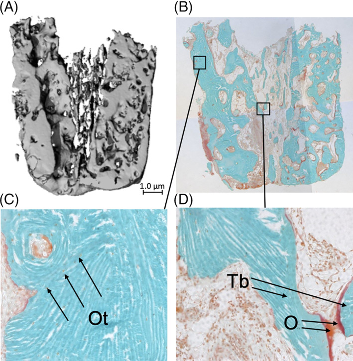
Histological and micro‐CT analysis of a biopsy taken from a double‐plated very thin knife‐edge alveolar ridge of an edentulous patient who received a reconstruction of the maxilla using calvarial bone grafts. A, Image of micro‐CT scan. Only bone with a mineral density of >559.2 mgHA/cm3 is visible. Magnification ×20. B, Histological section of the biopsy stained with Goldner's trichome to distinguish mineralized bone tissue (green) and unmineralized osteoid (red). Viable, mineralized mature bone(green) is visible. Osteoid is red. The morphology of the bone graft is still visible. Magnification ×20. C, Cortical region of interest (ROI), showing compact lamellar bone with several osteocytes visible as tiny black dots inside the green mineralized tissue, indicating vital bone. In the upper left corner, a haversian channel is visible. Magnification, ×100. Ot, osteocyte. D, The cancellous bone at the transition between grafted and native bone, the presence of osteoid (red; lower right corner) indicates the formation of new bone. Probably, the two trabeculae will be connected after maturation (mineralization) of the osteoid. Magnification, ×100. O, osteoid; Tb, trabecula
