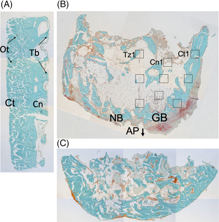
FIGURE 4.

Histological sections of biopsies obtained from one edentulous patient who received reconstruction of the maxilla using calvarial bone grafts: fresh calvarial bone biopsy and biopsies from the maxillary alveolar ridge, four and 45 months post‐reconstruction surgery. A, Fresh calvarial bone showing dense cortical bone (Ct) and several thick trabeculae (Tb) on the right, where the more cancellous diploic bone (Cn) starts. B, Biopsy 4 months after grafting with a native maxillary bone (NB; left), and grafted calvarial bone (GB; right). The cortical part of the calvarial bone is denser compared to the native bone. Between the native bone and calvarial grafted bone, crossing trabeculae are scarce, and non‐mineralized connective tissue is present. C, Biopsy 45 months after grafting. The border between grafted and native bone has disappeared, and there is more homogenous mineralized, hard bone tissue visible throughout the section compared to the section obtained after 4 months of graft healing. Staining: Goldner's trichome to distinguish mineralized bone tissue (green) and unmineralized osteoid (red). All bone/biopsies are from one patient, showing a progression from fresh calvarial bone toward a healed, reconstructed alveolar process. Magnification, ×20. AP, alveolar process; Cn, cancellous bone; Cn1, first ROI of the cancellous zone of grafted bone; Ct, cortical bone; Ct1, first ROI of the cortical zone of grafted bone; GB, grafted (calvarial) bone; NB, native (maxillary) bone; Ot, osteocyte; Tb, trabecula; Tz1, first ROI of the transition zone between grafted and native bone
