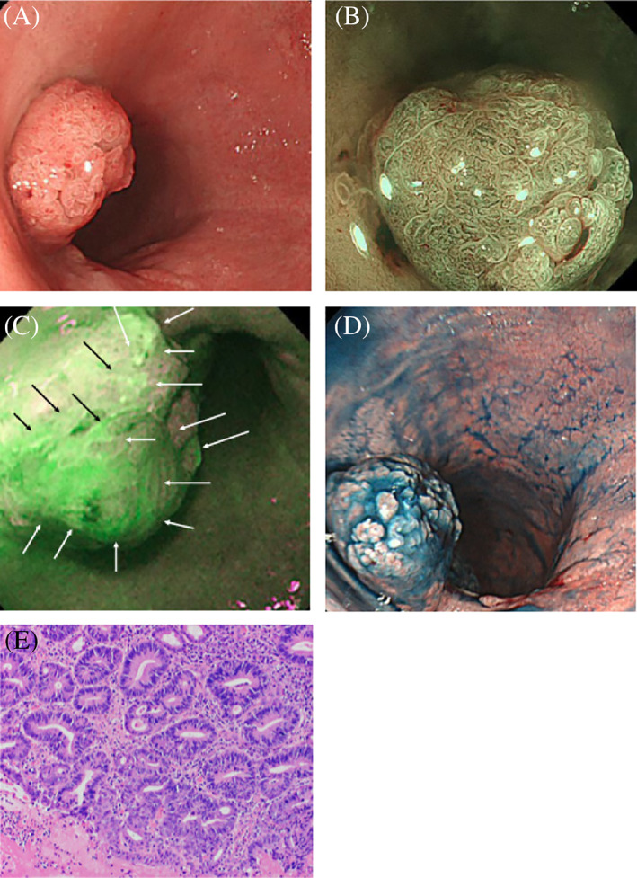
FIGURE 3.

A, White light endoscopy showing a protruding rectal lesion; B, Narrow‐band imaging combined with endoscopy showing a villous surface with irregular microvessels; C, Autofluorescence endoscopy showing a strong signal over the entire surface of the lesion (arrows); D, Pigment scatter image of the lesion; E, Histopathology showing an adenocarcinoma (HE stain, ×200)
