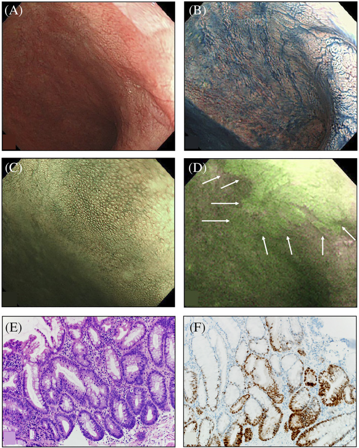
FIGURE 4.

A, White light endoscopy; B, Chromoendoscopy with indigo carmine; C, Narrow‐band imaging‐magnifying endoscopy does not show any lesions near the anal verge in the lower rectum; D, Autofluorescence endoscopy using 5‐aminolevulinic acid showing irregularly shaped areas with strong green fluorescent signals (arrows); E, Histopathology showing high‐grade dysplasia characterized by cellular and structural dysplasia (HE stain, ×200); F, Immunohistology showing p53‐positive epithelial cells (×200)
