Figure 1.
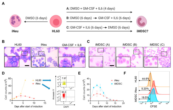
Induction of neutrophils and myeloid-derived suppressor cells (MDSCs) from HL60. (A) Experimental scheme for induced neutrophil (iNeu) and induced myeloid-derived suppressor cell (iMDSC) induction. Created with biorender.com. (B) Wright–Giemsa staining of HL60, iNeu, and HL60 treated with granulocyte macrophage-colony stimulating factor (GM-CSF) + interleukin 6 (IL6) for 6 days. Scale bar 50 μm. (C) Wright–Giemsa staining of iMDSC induced by the three conditions shown in (A). Scale bar 50 μm. (D) Proliferation curves and apoptosis analysis of HL60 and iNeu. Apoptotic cells were identified as Annexin V+ DAPI− cells. (E) Proliferation curves of iNeu and iMDSC. (F) Representative carboxyfluorescein succinimidyl ester (CFSE) flow cytometer histograms showing the differentiation dilution of the CFSE labeling of HL60, iNeu and iMDSC on Day 4 after the start of induction.

