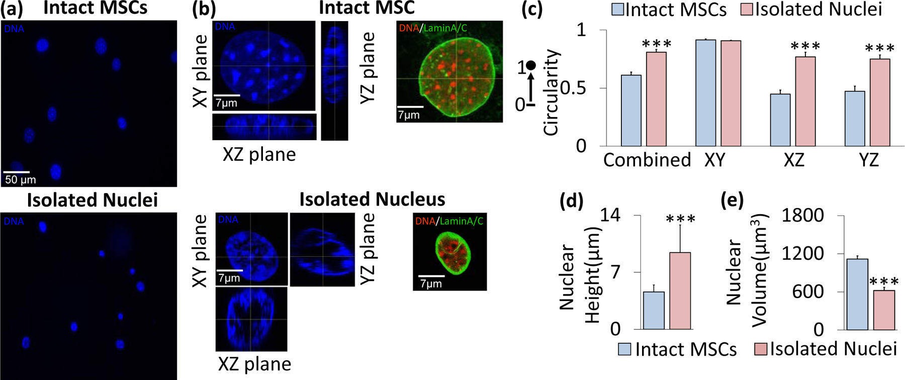
Figure 1. Nuclear geometry characteristics before and after nuclear isolation.

a) Mesenchymal stem cells (top) and isolated nuclei (bottom) were stained with Hoechst 33342 and subsequently imaged for DNA using a fluorescence microscope. b) Confocal imaging of an intact nucleus (top) and isolated nucleus (bottom) with Hoechst 33342 fluorescence staining under 63x focus with 16 Z-stacks for each nucleus image. Representative intact MSC (top) and isolated nucleus (bottom) shown in XY, XZ, and YZ planes of focus. Staining against LaminA/C also indicated an intact nuclear lamina post-isolation. c) Shape profiles of the intact MSC nuclei versus isolated nuclei. The isolated and intact nuclei exhibited similar circularity in the XY plane (i.e., the horizontal plane of the cell culture dish or microscope slide), but showed significant differences in shape profiles in the vertical XZ and YZ planes (p<.001, N=10 isolated nuclei, N=10 intact nuclei), with isolated nuclei showing 52% and 45% higher circularity values in those planes, respectively. The combined data, which is an average of all three planes, shows a significant difference in sphericity (p<.001) between the isolated nuclei and intact nuclei, with the two types having sphericity values of 0.809 and 0.612, respectively. d) Average isolated nuclear height (9.43 μm) is approximately twice that of intact MSC nuclei (4.59 μm, N=10/group, p<.001). e) Volume decreases from 1,116 μm3 to 621 μm3 following isolation (N=10/group, p<.001. * p<.05, ** p<.01, *** p<.001, against control.
