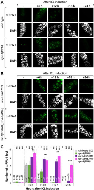
Figure 3. Formation and disappearance of RPA-1 foci after ICL induction are influenced by rev-1 mutation.
(A and B) L4 stage worms of wild type (N2), xpa-1(RNAi), rev-1(tm8701), and rev-1(tm8701); xpa-1(RNAi) were treated with TMP (200 μM) for 40 min and exposed to UV-A light (100 J/m2). Gonads were dissected, fixed, and immunostained using RPA-1 antibody at 0, 6, 12, 18 and 24 h after ICL induction. Scale bar, 10 μm. (C) The number of RPA-1 foci per nuclear focal plane was counted for over 100 mitotic germ cells in each stain. P values: *, P<0.05; **, P<0.01; ***, P<0.001; ns, no statistical significance. See Table S2 for One-way ANOVA analysis.

