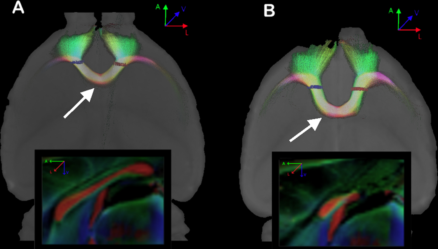
Figure 5. Probabilistic reconstruction of fibers connecting both frontal lobes.

In A, a Balb/c with a normal corpus callosum. In B, another Balb/c with alterations of the corpus callosum. Note the level where the frontal fibers cross the midline in both animals (arrows), and the difference between both CCs shown in the sagittal views (boxes). ROIs are shown in red and blue.
