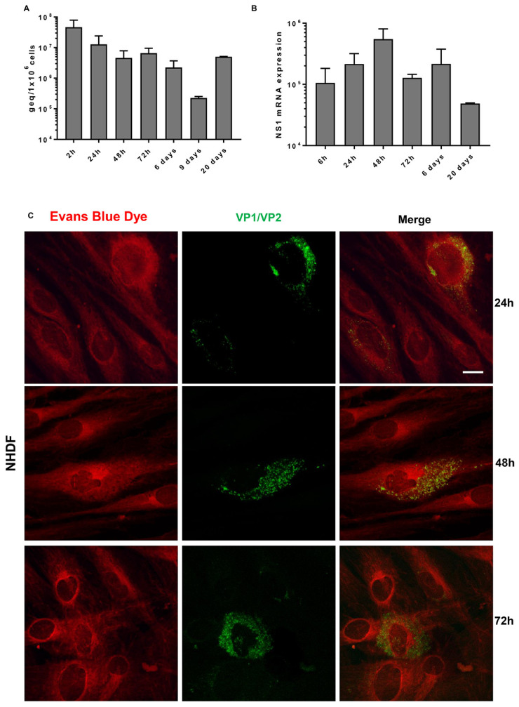
Fig. 1.
Time course of B19V infection of primary NHDF cultures
(A) The levels of viral DNA were evaluated at the end of absorption/penetration period (2 hpi), at 24, 48 and 72 h and at 6, 9 and 20 days post infection. (B) The mRNAs for NS1 protein were carried out at 6, 24, 48 and 72 h and at 6 and 20 days post infection. Bars represent mean (S.e.m.) values from four independent cultures. (C) Immunofluorescence assay for the detection of B19V VP1/VP2 proteins was carried out at 24, 48 and 72 hpi. Positive cells are stained in green. A counterstain with Evans Blue was performed in order to highlight the cell structure. Scale bar=5 µm.

